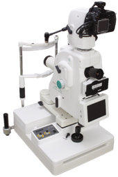

Over the years these camera filters, optical systems, and sensors have become more refined to maximize the fluorescence effect for clinical use. In the case of fluorescein angiography, the excitation wavelengths (that pass through the camera’s excitation filter) are in the ultraviolet/blue spectrum, and the emission wavelengths (that return through the camera’s barrier filter) are in the blue/yellow spectrum.
.jpg)
Fluorescein angiography (FA) requires intravenous injection of fluorescein sodium dye and uses special filters in the retinal camera to enhance the dye fluorescence.įluorescence consists of emission of longer wavelength of light or radiation from a substance as a result of a shorter wavelength light or radiation striking that substance. Since the introduction of human retinal fluorescein angiography in 1959 by Harold Novotny, MD, and David Alvis, MD, the ability to study, diagnose, and manage numerous retinal pathologies has expanded greatly. One of these technologies that has evolved over this period of time is fundus autofluorescence (FAF). Retinal imaging technology has taken great strides over the last several decades, and eyecare practitioners have the benefit of many different technologies to help diagnose and manage posterior segment eye disease. This article demonstrates the utility of using FAF technology to identify numerous fundus conditions and speculates on FAF use as a potential future tool for identifying SARS-CoV-2 infection in the eye. One of these technologies is fundus autofluorescence (FAF). ODs have several technologies available to assist in the diagnosis and management of patients’ ocular and visual conditions during the current novel coronavirus pandemic.


 0 kommentar(er)
